


- Medical Devices
- Confocal Microscope Market
Confocal Microscope Market Size, Share, and Growth Forecast, 2025 - 2032
Confocal Microscope Market By Product Type (Laser Scanning Microscopes, Spinning Disk Confocal Microscopes), Application (Life Sciences and Biomedical Research, Material Science and Nanotechnology), End-user, and Regional Analysis for 2025 - 2032
Confocal Microscope Market Size and Trends Analysis
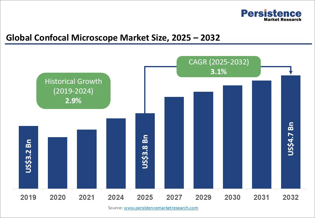
The global confocal microscope market size is likely to be valued at US$3.8 Billion in 2025 and is estimated to reach US$4.7 Billion in 2032, growing at a CAGR of 3.1% during the forecast period 2025 - 2032, driven by the rising demand for specialized applications across life sciences, material science, and nanotechnology. Developments such as the integration of circularly polarized light are expanding their scope.
Key Industry Highlights
- Leading Region: Asia Pacific, with about a 38.4% share in 2025, owing to investments in research and novel imaging infrastructure.
- Fastest-growing Region: North America, backed by ongoing technological developments and widespread clinical adoption.
- New Product Launch: Nikon Corporation released the AX R MP with NSPARC super-resolution multiphoton confocal microscope. It enables extremely sensitive array detection and super-resolution in deep areas within large living organism specimens.
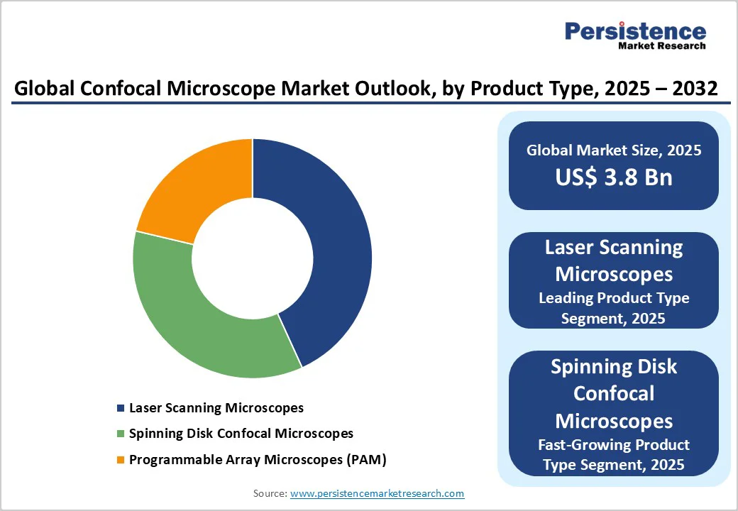
- Leading Product Type: Laser scanning microscopes hold nearly 43.2% share in 2025, spurred by high-resolution imaging and precise optical sectioning.
- Dominant Application: Life sciences and biomedical research, approximately 47.3% of the confocal microscope market share in 2025, as the microscopes are required for studying cellular structures.
- Key End-user: Diagnostic laboratories recorded about 35.6% share in 2025, because they require high-precision imaging for accurate molecular diagnostics.
| Key Insights | Details |
|---|---|
| Confocal Microscope Market Size (2025E) | US$3.8 Bn |
| Market Value Forecast (2032F) | US$4.7 Bn |
| Projected Growth (CAGR 2025 to 2032) | 3.1% |
| Historical Market Growth (CAGR 2019 to 2024) | 2.9% |
Confocal Microscope Market Size and Trends Analysis
Growth Analysis - Improvement of Imaging Precision and Automation
The growth of confocal microscopes is driven by developments in imaging precision and automation. Modern confocal systems now integrate high-speed laser scanning, multi-channel detection, and real-time image processing, enabling researchers to capture detailed three-dimensional structures with minimal manual intervention.
Automation features such as motorized stages and AI-assisted focusing reduce human error and accelerate data acquisition, making experiments more reproducible. These improvements are important in live-cell imaging, where maintaining cell viability is essential. For instance, the Leica TCS SP8 system combines automated image acquisition with high-resolution scanning, allowing researchers to monitor dynamic cellular events efficiently.
Expansion into Specialized Research Applications
Another key growth driver is the increasing use of confocal microscopes in specialized scientific fields. Researchers are applying confocal imaging to areas such as neuroscience, nanotechnology, and pharmacology, where precise visualization of micro- and nanoscale structures is essential.
The ability to customize optical configurations and integrate additional techniques such as Raman spectroscopy or multiphoton imaging allows scientists to investigate highly specific biological, chemical, or material phenomena. For example, the University of Cambridge uses confocal Raman microscopy to study 2D materials at the nanoscale, revealing properties important for electronic and mechanical applications.
Barrier Analysis - Risk of Cellular Damage from High-intensity Lasers
One major restraint to the adoption of confocal microscopes is the potential harm caused to living cells by high-intensity laser exposure. Prolonged or intense illumination can induce heat and generate reactive oxygen species, leading to phototoxicity and cellular stress. This limits the duration and frequency of live-cell imaging experiments as researchers must carefully balance image quality with cell viability.
For instance, in neurobiology studies, high-resolution imaging of neurons over extended periods can result in cellular damage if laser intensity is not carefully controlled. Such constraints require the use of novel techniques, including spinning disk confocal systems or lower-intensity multiphoton imaging to mitigate damage, adding complexity and cost to experiments.
Limited Excitation Wavelength Options in Standard Lasers
Another challenge is the narrow range of excitation wavelengths provided by commonly used lasers. Several traditional laser sources, such as argon or helium-neon lasers, only cover specific portions of the visible spectrum, limiting the number and type of fluorophores that can be simultaneously imaged. This constraint reduces flexibility in multi-label experiments, which are often required in complex biological studies.
For example, researchers investigating multiple protein interactions in cancer cells tend to find standard laser configurations inadequate for detecting all relevant fluorophores. Overcoming this limitation often requires additional or tunable laser sources, increasing the complexity and cost of the microscopy setup.
Opportunity Analysis - Spectral Imaging and Fluorescence Unmixing
A key growth opportunity for confocal microscopes lies in spectral imaging combined with fluorescence unmixing. This technology allows researchers to distinguish overlapping fluorescent signals in complex biological samples, enabling simultaneous imaging of multiple biomarkers with high specificity. This approach is useful in studies involving heterogeneous tissues or multi-protein interactions, where conventional imaging would struggle to differentiate signals.
For example, the Zeiss LSM 980 with Airyscan 2 employs spectral detection and unmixing to visualize multiple fluorescent proteins in live-cell imaging, providing clear insights into cellular dynamics. The adoption of spectral microscopy improves data accuracy, reduces experimental repetition, and opens new avenues in advanced biomedical research.
Opportunities in Circularly Polarized Light Applications
The use of Circularly Polarized Light (CPL) in confocal microscopy presents another promising growth area. CPL enables the study of chiral molecules and materials, providing unique insights into molecular orientation, structural asymmetry, and optical activity at micro- and nanoscale levels. This capability is valuable in fields such as material science, pharmaceuticals, and biomolecular research, where understanding chirality is important.
For instance, researchers at ETH Zurich have used CPL-integrated confocal systems to examine protein folding patterns and asymmetric crystal growth, revealing subtle structural features that conventional microscopy could not detect. By integrating circularly polarized light, confocal microscopes can serve highly specialized research applications.
Category-wise Analysis
Product Type Insights
Laser scanning microscopes are anticipated to account for nearly 43.2% of the share in 2025. This is due to their ability to provide exceptional image resolution and optical sectioning, allowing researchers to capture detailed 3D structures of cells and tissues.
They are valuable in applications requiring precise localization of molecules, such as neuroscience studies and live-cell imaging. For example, Nikon’s A1R HD25 laser scanning microscope is widely used in cancer research for visualizing tumor microenvironments with high clarity and accuracy.
Spinning disk confocal microscopes recorded around 26.8% of the share in 2025, owing to their ability to perform high-speed imaging with minimal phototoxicity, making them ideal for live-cell experiments. They allow fast acquisition of multiple focal planes simultaneously, which is essential for observing dynamic processes, including intracellular transport and neuronal activity.
Olympus’s IXplore SpinSR system, for instance, has gained popularity for real-time imaging of live zebrafish embryos and other fast biological processes.
Application Insights
The life sciences and biomedical research segment is poised to account for approximately 47.3% of the share in 2025. Confocal microscopes allow precise imaging of cellular and subcellular structures, which is essential for understanding disease mechanisms, drug interactions, and genetic studies.
For example, researchers at Harvard Medical School use confocal microscopy to study neuronal networks and track protein localization in live cells, providing key insights for neurodegenerative disease research.
The material science and nanotechnology segment is seeing decent growth as confocal microscopes enable high-resolution surface and structural analysis at the micro- and nanoscale. This capability is important for developing advanced materials, semiconductors, and nanodevices. For instance, the University of Cambridge uses confocal Raman microscopy to analyze nanoscale features in 2D materials such as graphene.
End-user Insights
Diagnostic laboratories are expected to hold a share of about 35.6% in 2025, as they require high-resolution imaging to accurately detect pathogens, analyze tissue samples, and perform molecular diagnostics. Confocal systems enable detailed visualization of cellular structures, which improves the reliability of test results.
Hospitals are a significant end-user, as confocal microscopes support novel clinical diagnostics and research directly linked to patient care. They are used for real-time imaging in dermatology, ophthalmology, and oncology, allowing clinicians to monitor disease progression and guide treatment decisions.
Regional Insights
Asia Pacific Confocal Microscope Market Trends
Asia Pacific is estimated to account for nearly 38.4% of the share in 2025, owing to ongoing research activities and technological developments. China, Japan, and India are at the forefront, with China leading in market share due to its substantial investments in research and development. Technological development is a key driver in Asia Pacific.
For instance, the development of sophisticated and user-friendly confocal Raman systems is improving the capabilities of confocal microscopy in material science and life sciences. The market is also witnessing a shift toward modular and customizable confocal systems, allowing researchers to customize instruments to specific requirements, thereby increasing accessibility and application across various scientific disciplines.
North America Confocal Microscope Market Trends
In North America, confocal microscopy is experiencing steady growth, specifically in the U.S. and Canada. The market is projected to reach approximately US$580 Million by 2032, propelled by developments in live-cell imaging and applications in fields such as dentistry, pharmaceuticals, and diagnostics. The adoption of modular confocal systems is increasing, providing customizable configurations to meet specific research requirements.
Technological developments are at the forefront of the regional market growth. Institutions such as Rensselaer Polytechnic Institute and Albany Medical College are sharing novel imaging equipment, including rare microscopes critical for biomedical research. For instance, Albany Medical College recently acquired a Nikon AXR MP multiphoton microscope. It helped in reducing imaging time from hours to minutes, enabling real-time cellular analysis essential for studying rapid biological interactions.
Europe Confocal Microscope Market Trends
In Europe, confocal microscopy is integral to advanced research and clinical diagnostics. Institutions such as the European Molecular Biology Laboratory (EMBL) and the Institute of Cancer Research (ICR) in London utilize confocal systems for high-resolution imaging in cell biology and oncology studies. These facilities often employ laser scanning and spinning disk confocal microscopes to observe dynamic cellular processes and tumor microenvironments in real-time.
Technological development is a hallmark of European confocal microscopy. For example, the Italian Institute of Technology in Genoa developed a compact microscope equipped with a Single-Photon Avalanche Diode (SPAD) array detector, enabling high-resolution structural and functional imaging in a single architecture. This development improves imaging capabilities for various biological applications.

Competitive Landscape
The global confocal microscope market is characterized by the presence of several established players, each providing a range of products catering to various applications in research and industry. Companies such as Carl Zeiss AG, Leica Microsystems (Danaher Corporation), Nikon Corporation, Olympus Corporation, and Thorlabs, Inc. are prominent in this market, providing novel imaging solutions for applications in the life sciences, material sciences, and semiconductor industries.
Key Industry Developments
- In May 2025, Evident launched two new spinning disk confocal microscopes, the IXplore IX85 SpinXL and the IXplore IX85 SpinSR. These added powerful new technology to the platform, broadening the possibilities of what researchers can capture and discover in live-cell imaging.
- In March 2025, Zeiss introduced Lightfield 4D, a new microscopy system based on the light-field principle. It is expected to change the way researchers observe living organisms, especially in neurosciences, cancer research, developmental biology, and plant sciences.
Companies Covered in Confocal Microscope Market
- Olympus Corporation
- Nikon Corporation
- ZEISS Group (Carl Zeiss Meditec AG)
- Danaher Corporation (Leica Microsystem)
- Agilient Technologies
- Bruker Corporation
- Caliber Imaging & Diagnostics
- Horiba
- ISS Inc.
- Confocal.nl
- Thorlabs Inc.
- Oxford Instruments PLC
Frequently Asked Questions
The confocal microscope market size is projected to reach US$3.8 Billion in 2025.
Advances in imaging resolution and automation are the key market drivers.
The confocal microscope market is poised to witness a CAGR of 3.1% from 2025 to 2032.
The rising adoption of modular confocal systems and the integration of fluorescence unmixing are the key market opportunities.
Olympus Corporation, Nikon Corporation, and ZEISS Group (Carl Zeiss Meditec AG) are a few key market players.


